Guides and Protocols
Our laboratory uses standardized protocols for the collection, preservation, extraction, and quantification of nucleic acids, specifically optimized for water hyacinth (Pontederia crassipes).
Two distinct sampling strategies are considered according to the biogeographic status of the population: (1) populations occurring in invaded areas and (2) populations occurring within the native range of P. crassipes. These scenarios differ primarily in the spatial scale at which sampling is designed.
The procedures described below provide a common framework for collaborators. They may be adapted according to site accessibility, availability of plant material, collection permits, local regulations, field conditions, laboratory infrastructure, available equipment, storage capacity, and sequencing-provider requirements. When a recommendation cannot be followed, retaining clear metadata and documenting any relevant methodological differences is encouraged.
🌿 1. Tissue Collection and Herborization
Sampling Design
Sampling in Invaded Areas
In invaded regions, P. crassipes populations commonly occur in relatively delimited inland freshwater systems such as lakes, lagoons, wetlands, reservoirs, ponds, and other enclosed or semi-enclosed water bodies.
As a reference sampling design for each site or population:
- Aim to collect samples from three individual plants.
- Select individuals as far apart from one another as the site and accessibility allow.
- Because P. crassipes reproduces extensively through clonal propagation, avoid visibly interconnected ramets or individuals belonging to the same compact clonal mat whenever possible.
- From each individual, reserve approximately 3–5 healthy leaves, depending on tissue availability.
- Keep tissue collected from different individuals separate whenever possible and assign a unique sample code to each individual.
- If site size, accessibility, population size, permits, or other field conditions limit the number or spatial separation of samples, collect the material that can reasonably be obtained and document the circumstances under fieldNotes.
The reference design therefore considers three spatially separated individuals per site, while recognizing that the effective number of samples may vary according to local conditions.
Sampling in the Native Range
Within the native range of P. crassipes, particularly in rivers, floodplains, and connected aquatic systems of the Amazon and other South American hydrological basins, sampling can be designed to represent the basin or connected hydrological system, rather than concentrating exclusively on a single locality.
This basin-level sampling framework is consistent with previous population genomic sampling of water hyacinth across hydrological basins within its native range (da Cunha et al., 2022).
As a reference design for native-range sampling:
- When feasible, distribute sampling among multiple points along the river, basin, or connected hydrological system.
- Where a clear flow direction or longitudinal hydrological gradient exists, sampling points can represent upstream, intermediate, and downstream portions of the system. This is an operational criterion adopted by the initiative to improve spatial representation of the basin.
- Where no clear upstream–downstream gradient is present, sampling points can instead be distributed as broadly as practical throughout the accessible portion of the basin or aquatic system.
- At each sampling point, aim to collect samples from three individual plants.
- Select the individuals at each point as far apart as local conditions allow, avoiding visibly interconnected ramets whenever possible.
- From each individual, reserve approximately 3–5 healthy leaves, depending on tissue availability.
- Keep each individual as an independent sample whenever practical and assign it a unique sample code.
- Additional sampling points along the basin are encouraged when access, logistics, permits, and available resources allow.
- The relative position of each sampling point within the basin or river system can be recorded under fieldNotes.
Under this reference design, three individuals correspond to each sampling point rather than to the entire basin. However, the number of sampling points and individuals can be adjusted according to the scale of the system and the practical capacity of each collaborator.
Tissue Collection for DNA Extraction
For optimal DNA preservation during field sampling and subsequent transport or storage, silica-gel drying is recommended as soon as possible after collection.
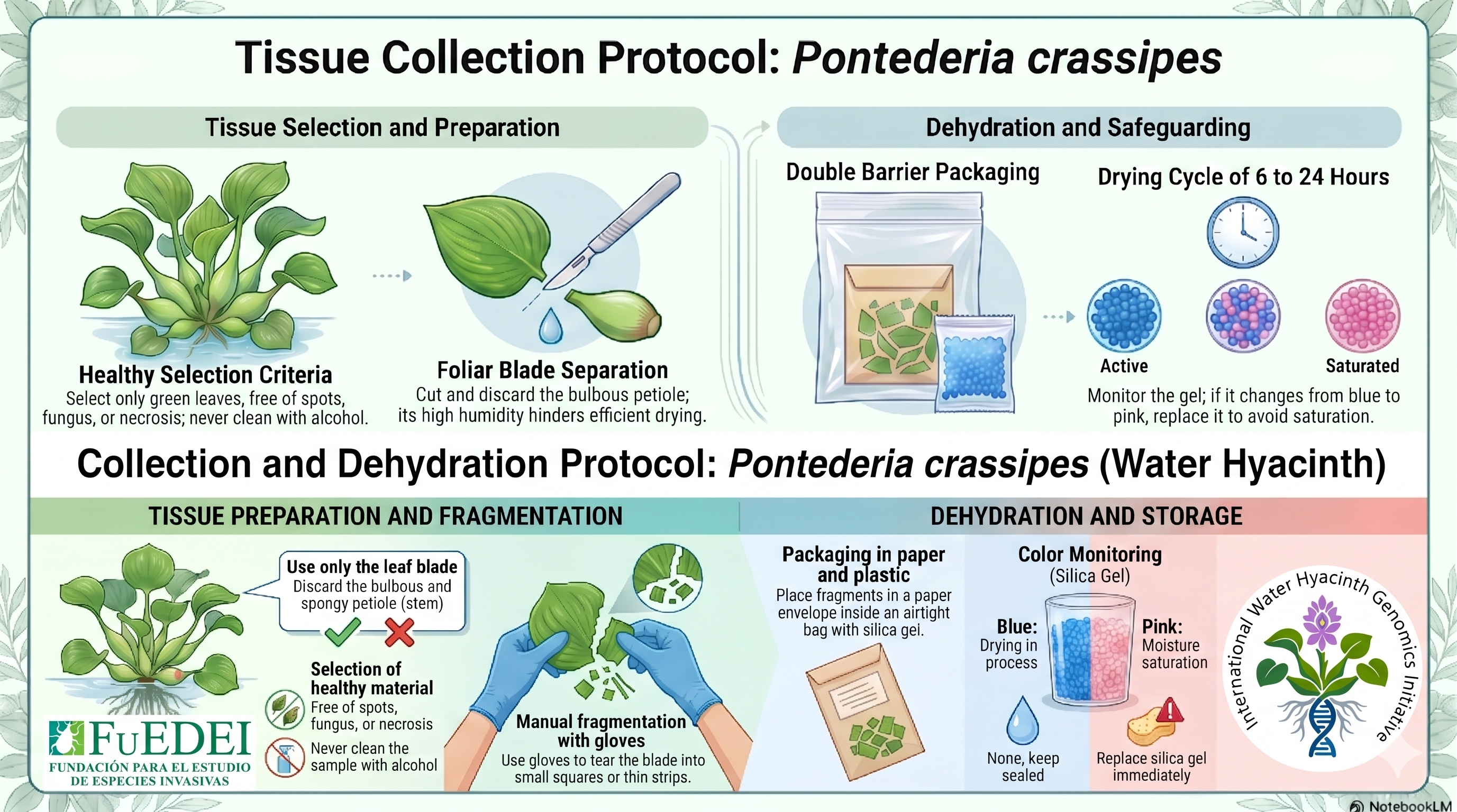
- Select healthy, preferably young leaves of P. crassipes.
- Reserve approximately 3–5 leaves from each sampled individual, according to tissue availability.
- Cut representative portions of the leaves into smaller pieces, avoiding excessively thick, highly hydrated, or spongy tissues whenever possible.
- Place tissue from each individual in a separate, clearly labeled zip-lock bag containing color-indicating silica gel whenever sufficient material and supplies are available.
- Keeping tissue from different individuals separate is recommended to preserve individual-level information.
- Use enough silica gel to promote rapid dehydration of the plant material.
- Allow the tissue to dry thoroughly. Well-dried tissue generally becomes brittle and snaps when bent.
- Replace or regenerate silica gel when the color indicator shows substantial moisture.
Fresh material may also be processed when DNA extraction can be performed locally shortly after collection and an appropriate cold chain is available. Silica-gel drying is the preferred preservation method for samples that will be transported or stored before DNA extraction, but alternative preservation strategies may be used when they are more appropriate for local conditions.
Photographic Documentation
Photographic documentation is strongly recommended for sampling sites and collected material and is particularly valuable for samples that are not directly represented by a herbarium voucher.
Whenever possible, collaborators are encouraged to archive:
- A general photograph of the sampled population and sampling site, showing the water body and surrounding environmental context.
- Photographs of sampled or representative P. crassipes individuals in situ, preferably showing diagnostic vegetative and reproductive structures when available.
- Individual-level photographs linked to the DNA sample code, particularly for samples that will not have a directly associated voucher specimen.
- A photograph of each voucher or pressed specimen that is prepared.
Additional photographs documenting population density, associated vegetation, habitat characteristics, flowers, fruits, environmental disturbances, or other relevant features can provide valuable supporting information.
Whenever possible, photographs should be associated with the corresponding locality, sampling point, and sample codes so they can be linked to the global sample database.
For DNA samples without an individual herbarium voucher, photographic documentation becomes especially important as supporting evidence of the identity, condition, and provenance of the sampled material.
Herborization and Voucher Specimens
It is not necessary to prepare a herbarium voucher for every individual sampled for DNA. Voucher collection is intended to provide representative and verifiable botanical documentation of the sampled populations without generating unnecessary duplication of herbarium material.
Voucher preparation can be prioritized according to the characteristics and spatial structure of the sampling:
- Invaded areas: when feasible, prepare a representative voucher for each distinct sampling site, population, or water body. If several sampling points belong to the same continuous and morphologically homogeneous population, a single well-documented voucher may be sufficient to represent them.
- Native range: representative vouchers can be prepared for distinct localities, hydrological sectors, or populations across the sampled basin. It is generally unnecessary to prepare a separate voucher for each of the three DNA samples collected at a sampling point.
- Additional vouchers are especially useful when there is noticeable morphological variation, taxonomic uncertainty, substantial geographic separation, contrasting habitat conditions, or reproductive material that adds diagnostic information.
- When available, flowering or fruiting material is particularly useful for taxonomic documentation.
- Voucher information can be cross-referenced with the DNA sample codes collected from the same population, locality, or sampling point.
The objective of herborization is therefore to provide representative taxonomic documentation of the sampled populations rather than to duplicate every DNA sample as a herbarium specimen.
When logistical conditions, permits, available material, herbarium capacity, or other factors make voucher preparation impractical, high-quality photographic documentation and complete associated metadata provide an important alternative source of supporting information.
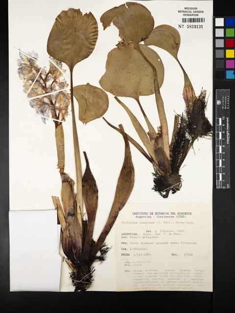
Follow the specific herbarium protocols of your local institution when available. If no specific guidelines for aquatic plants are provided by your local herbarium, the following method adapted for aquatic flora (Rodríguez & Rojas, 2006) can be used:
- Aquatic Plant Pressing: Avoid pressing directly into dry newspaper when delicate aquatic parts, such as Pontederia flowers, could adhere to the paper and be damaged upon removal.
- Collect the plants in wet newspaper and roll them up.
- In the laboratory/herbarium, place the plant in a tray of water to allow the structures to spread and extend naturally.
- Slide a piece of white cardstock under the plant and carefully lift it out of the water, keeping the plant positioned on the cardstock.
- Place the cardstock with the plant inside the pressing paper (newspaper/blotters) and proceed with the drying method normally used by the herbarium.
Sample Metadata
Consistent sample metadata are highly valuable for integrating records into the global database. Collaborators are encouraged to record as many of the following fields as possible for each collected sample.
Some fields may not be available for every sample. Missing information can be recorded as NA when appropriate rather than preventing the sample from being incorporated into the dataset.
For samples from the native range, the relative location of the sampling point within the hydrological system, such as upstream, intermediate, downstream, or another relevant spatial description, can also be recorded under fieldNotes.
Please use these fields whenever the corresponding information is available:
| Field Name | Description & Examples |
|---|---|
| code | Unique sample identifier (e.g., CAM0001, M04) |
| no_samples | Number of samples collected (e.g., 1, 3, 5) |
| preservation | Tissue preservation method (e.g., Dry leaf in silica, Fresh leaf) |
| herbo | Herbarium code and/or voucher identifier associated with the sampled population or locality, when available; use NA if no voucher was prepared |
| country | Country of collection (e.g., Argentina, Mexico) |
| stateProvince | State, province, or major region (e.g., Buenos Aires) |
| locality | Specific collection site (e.g., Otamendi, Laguna San Vicente) |
| decimalLatitude | Latitude in decimal degrees (e.g., 12.020736) |
| decimalLongitude | Longitude in decimal degrees (e.g., -61.762672) |
| elevation | Elevation in meters above sea level |
| waterBody | Name or type of the water body |
| Date (year/month/day) | Year, Month, and Day of collection |
| fieldNotes | Relevant observations about the plant, population, environment, position within the basin, voucher association, or sampling limitations |
| recordedBy | Name of the collector (e.g., A. Faltlhauser, A. Sosa) |
| scientificName | Pontederia crassipes |
| degreeOfEstablishment | Status in the area (e.g., Native, Invasive) |
🧬 2. DNA Extraction
DNeasy® Plant Pro Kit (QIAGEN)
The DNeasy® Plant Pro Kit (QIAGEN) is the DNA extraction kit used, tested, and validated in our laboratory for Pontederia crassipes. The protocol described below corresponds to the workflow currently used by our laboratory.
The reagent conditions indicated below follow the manufacturer’s recommendations for plant tissues rich in phenolic compounds and other secondary metabolites. They should therefore not be interpreted as an independent modification developed by the initiative.
The specific brand or model of tissue disruption equipment may vary among collaborating laboratories. The objective is to obtain adequate tissue pulverization using the equipment locally available.
Starting Material & Pre-treatment
- Silica-gel dried leaf tissue is the standard starting material used in our workflow for samples collected, transported, or stored before extraction.
- Fresh or recently collected tissue can also be processed when adequate cold storage and rapid extraction are available.
- Immediately before disruption, freezing the tissue with liquid nitrogen is recommended when available. If liquid nitrogen is not accessible, overnight storage at -20°C or -80°C can be used as an alternative, or another validated procedure suitable for the collaborating laboratory can be followed.
Preparation and Homogenization
- Transfer the frozen tissue to the appropriate disruption tube.
- Lysis Buffer: Use approximately 5–100 mg of tissue in a 2 ml tissue disruption tube.
- Following the DNeasy® Plant Pro Kit recommendations for plant material with high phenolic content, add 450 µl of Solution CD1 and 50 µl of Solution PS.
- Vortex briefly to mix.
- Homogenization: Any suitable tissue disruption system available in the collaborating laboratory can be used, including a vortex with an appropriate adapter, bead beater, or tissue homogenizer.
- In our workflow, samples are vortexed at the maximum operational setting for approximately 3–5 minutes, with the objective of achieving thorough tissue disruption.
Extraction Steps
- Centrifuge the disruption tubes at 12,000 x g for 2 min.
- Transfer the supernatant to a clean 1.5 ml tube.
- Add 200 µl of Solution CD2 and vortex for 5 s.
- Centrifuge at 12,000 x g for 1 min.
- Avoiding the pellet, transfer the supernatant to a clean 1.5 ml tube.
- Add 500 µl of Buffer APP and vortex for 5 s.
- Load 600 µl of lysate onto an MB Spin Column.
- Centrifuge at 12,000 x g for 1 min and discard the flow-through. Repeat if necessary to process the entire lysate volume.
- Place the column into a clean 2 ml collection tube.
- Add 650 µl of Buffer AW1, centrifuge at 12,000 x g for 1 min, and discard the flow-through.
- Add 650 µl of Buffer AW2, centrifuge at 12,000 x g for 1 min, and discard the flow-through.
- Centrifuge the column at up to 16,000 x g for 2 min to dry the membrane.
- Elution: Add 50–100 µl of Buffer EB directly to the center of the membrane, allow it to sit briefly at room temperature, and centrifuge at 12,000 x g for 1 min.
RNase Treatment
The sequencing provider currently used by the initiative requests RNase-treated genomic DNA. For samples intended for this provider, RNase treatment can therefore be incorporated during extraction or as part of a post-extraction cleanup workflow.
- RNase A or another DNase-free RNase compatible with genomic DNA purification can be used.
- The amount of enzyme, incubation temperature, and incubation time can follow the instructions of the RNase product available in the collaborating laboratory.
- If RNase treatment is performed after DNA elution, any subsequent cleanup recommended by the RNase manufacturer or sequencing provider can also be performed.
- Collaborators using a different sequencing facility can follow the specific RNA-removal requirements established by that provider.
Because RNase formulations differ among manufacturers, a universal RNase volume is not specified here.
💧 3. DNA Quantification & Quality Control
Collaborators may use the spectrophotometer or fluorometer available in their facilities. Because P. crassipes contains substantial quantities of secondary metabolites, spectrophotometric and fluorometric measurements provide different types of information and are best interpreted accordingly.
Quality Assessment: Spectrophotometry (e.g., NanoDrop)
Spectrophotometers measure absorbance and may be affected by phytochemicals, carbohydrates, phenols, RNA, and other contaminants commonly encountered in plant DNA extractions. For this reason, absorbance measurements are particularly useful for assessing DNA purity and quality rather than absolute DNA concentration.
- A260/280 Ratio: Values around 1.8–2.0 generally indicate relatively pure DNA.
- A260/230 Ratio: Values around 1.8–2.2 are commonly associated with relatively clean DNA. In plant samples containing high concentrations of secondary metabolites, this ratio may be lower. Values substantially below 1.5 can indicate contamination that may affect downstream PCR or NGS applications (Desjardins et al., 2009).
These ranges are useful reference values rather than strict exclusion criteria, particularly for plant material that can be difficult to extract.
Quantity Assessment: Fluorometry (e.g., Qubit, DeNovix FX)
For accurate estimation of DNA concentration, fluorometric methods are preferred whenever they are available.
Fluorometric assays use dyes with greater specificity for double-stranded DNA (dsDNA) and are therefore less affected by the absorbance interference caused by plant secondary metabolites.
- When possible, report the final DNA concentration in ng/µl based on fluorometric measurements.
- Fluorometric concentration is preferred for calculating the amount of DNA available for genomic sequencing.
If fluorometric equipment is not locally available, collaborators can report the measurements available to them and indicate the quantification method used.
Approximate DNA Concentrations Observed in Our Laboratory
The following concentrations are provided only as approximate empirical reference values obtained during our work with P. crassipes. They are not acceptance thresholds and should not be interpreted as guaranteed extraction yields.
- Fresh or recently collected material: approximately 20–30 ng/µl.
- Material stored for longer periods in silica gel: approximately 4–8 ng/µl.
Lower concentrations may also occur and can still provide useful material depending on the sequencing strategy and provider requirements.
DNA recovery varies according to tissue condition, storage duration, silica-gel efficiency, initial tissue quality, tissue quantity, homogenization efficiency, extraction performance, and other sample-specific factors.
Whenever possible, individual quantification of each DNA sample is preferred over assuming concentration from tissue condition or preservation time.
🧬 4. Genomic Sequencing Guidelines
Collaborators may use any sequencing facility available to them and are encouraged to follow the sample-submission parameters established by their selected sequencing provider.
Sequencing depth can be adjusted according to the project objectives, sequencing strategy, available budget, laboratory resources, and material available from each sample.
When a sequencing facility does not provide more specific submission guidelines, the following values can be used as general reference targets:
- Total Amount & Quality: Approximately 100 ng of high molecular weight (>20 kb) genomic DNA is a useful target when sufficient material is available. Lower amounts may also be accepted depending on the library-preparation method and sequencing provider.
- RNase Treatment: For the sequencing provider currently used by the initiative, RNase-treated DNA is requested. Other providers may have different requirements.
- Target Concentration: Approximately 5.0–10.0 ng/µl is a useful working range. Depending on the sequencing provider and library-preparation workflow, samples outside this range may also be suitable.
- Quantification Method: Qubit, PicoGreen, DeNovix FX, or another dsDNA-specific fluorometric assay is preferred when available.
- Purity Assessment: Spectrophotometric measurements can provide complementary information on DNA purity and potential contamination.
- Final QC: Before shipment or sequencing submission, it is useful to review DNA concentration, purity, integrity when this can be evaluated, RNase-treatment status when applicable, and any additional requirements established by the selected sequencing facility.
Provider-specific requirements should take precedence over these general reference values.
🧬 5. Spatial Sampling Strategies
The sampling framework used by the initiative follows two clearly differentiated spatial strategies:
Invaded areas: sampling site or water body → aim for 3 spatially separated individuals → approximately 3–5 leaves per individual, depending on availability.
Native range: basin or connected hydrological system → multiple sampling points distributed along the system when feasible → aim for 3 spatially separated individuals per sampling point → approximately 3–5 leaves per individual, depending on availability.
These numbers represent the reference sampling design rather than fixed thresholds. Fewer individuals, fewer sampling points, or smaller amounts of tissue can still be incorporated when access, permits, population size, logistics, available resources, or other local conditions limit sampling.
The basin-level sampling approach for populations within the native range is consistent with previous population genomic sampling of P. crassipes across South American hydrological basins (da Cunha et al., 2022).
Reference
da Cunha, N. L., Xue, H., Wright, S. I., & Barrett, S. C. H. (2022). Genetic variation and clonal diversity in floating aquatic plants: Comparative genomic analysis of water hyacinth species in their native range. Molecular Ecology, 31, 5307–5325. https://doi.org/10.1111/mec.16664